fNIRSBrain imaging
Imaging a Baby’s Whole Brain with a Single Cap
High-density optical tomography (HD-DOT) has mapped brain activity across the entire scalp of 5–7-month-old infants — without ever going near an MRI scanner.
Infancy is when the brain develops fastest. So how do we image that developing brain without putting the baby inside a machine, while they play or look at a screen? This study looks for the answer in a wearable optical cap.
The method first: what are fNIRS and HD-DOT?
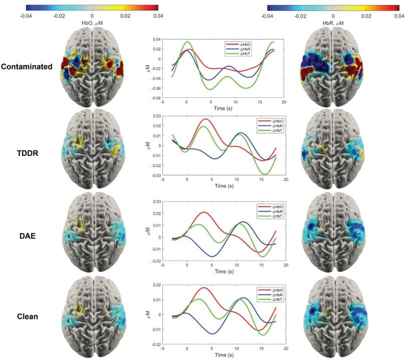
Functional near-infrared spectroscopy (fNIRS) and its high-resolution form, high-density diffuse optical tomography (HD-DOT), work with harmless near-infrared light sent in through the scalp. They measure changes in oxygenated and deoxygenated haemoglobin in the cortex, an indirect indicator of how active that region is. The greatest advantage: it is wearable and portable, and unlike MRI it does not require keeping still. That makes it ideal for babies.
The problem solved: the whole, not a part
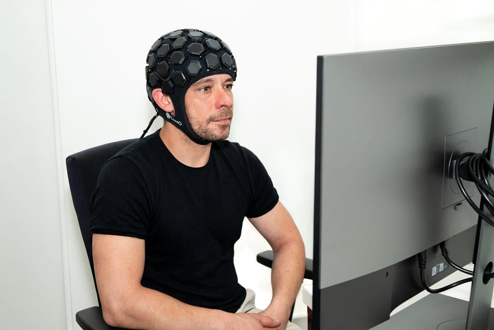
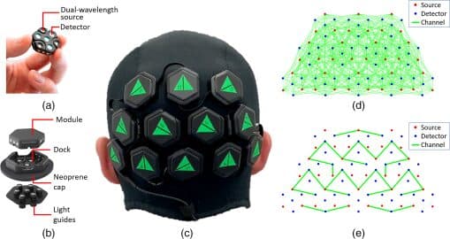
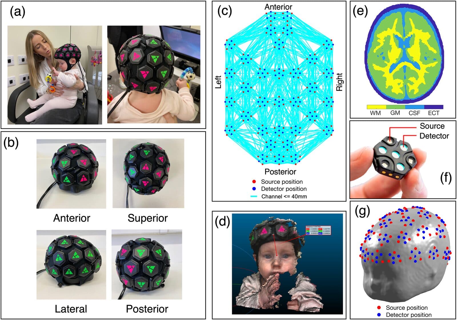
Earlier wearable caps shared one limitation: they could sample only a particular region of the cortex. The researchers developed a cap, built on the high-density modular LUMO design by Gowerlabs (UK), that can cover the baby’s entire scalp.
How was the study done?
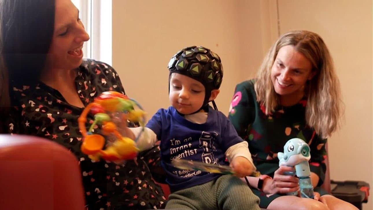
Recordings were taken from 16 infants aged 5–7 months while social and non-social audio-visual stimuli were shown on a screen. The aim was to see brain activity related to social processing with whole-head coverage.
The result, and why it matters
The study showed that activity can be mapped across the entire optically accessible cortex in infants — a proof of concept that goes far beyond the coverage of earlier methods. Whole-head imaging makes it possible to study processes that span several regions, such as social brain networks, in one go, and lays the groundwork for complex stimuli and advanced analyses such as decoding in the future.