fNIRSAdult / vision
Wearable Brain Imaging Rivals Giant Laboratory Systems
Wearable high-density optical tomography (HD-DOT) has proved it can map the adult visual cortex with the same quality as huge fibre-optic systems.
Brain imaging has long faced a dilemma: the highest-resolution optical methods were possible only with huge, unwieldy devices. This study shows that a wearable system can do the same job as those giant systems.
The problem: quality, but no portability
High-density optical tomography (HD-DOT) can reach a resolution and localisation accuracy in the adult brain close to that of fMRI (blood-oxygen-level-dependent MRI). It achieves this by scanning the tissue with dense, overlapping measurements. But until now this performance required large, unwieldy fibre-optic arrays — in practice, the device was confined to the lab.
Aim and method
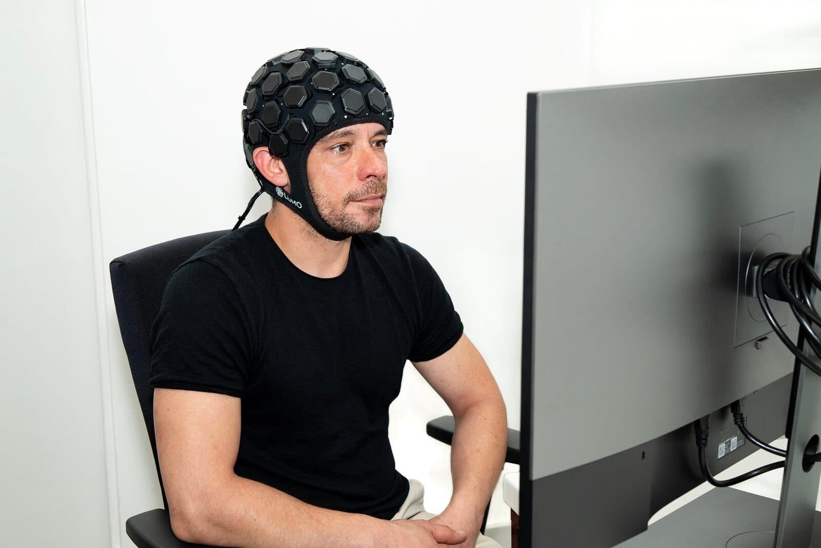
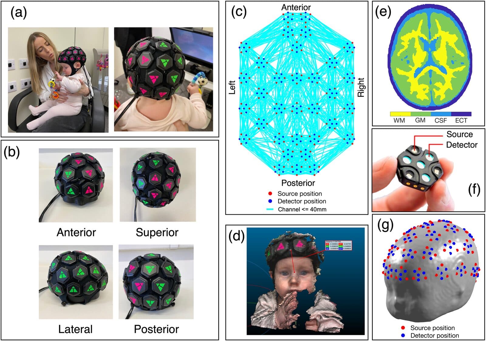
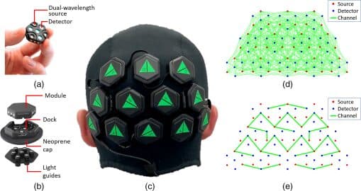
The researchers evaluated a wearable HD-DOT system (Gowerlabs LUMO) that provides the same sampling density as large fibre systems but is far more ergonomic. To do so, they reapplied classic visual stimulation paradigms — retinotopic mapping. By taking intensive measurements from a single person over 15 sessions, they tested both imaging performance and repeatability.
Retinotopy, in short, is this: positions in the visual field form an orderly “map” in the visual cortex. Recovering that order correctly is the classic way to test the quality of an imaging system.
Result
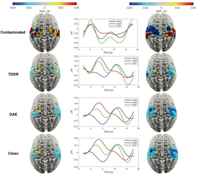
The haemodynamic response functions and cortical activation maps obtained with the wearable system reproduced the results obtained with large fibre-based systems. In other words, wearable HD-DOT delivered the same quality as giant tabletop systems.
Why does it matter?
This result is a major step in taking high-quality brain imaging out of the fixed laboratory and the MRI room. More comfortable, more natural and repeatable measurements mean far wider use, from the clinic to everyday research.