EEGEEG research
Mapping the Central Sulcus with the g.Pangolin Ultra-High-Density EEG/EMG/ECG System
Understanding how the brain works is extremely important. One of the most common ways to do so is to map brain activity with electroencephalography (EEG). EEG is easy, inexpensive and needs no surgery. In our latest publication, we set out to take brain mapping far beyond conventional methods with the g.Pangolin Ultra-High-Density EEG (uHD EEG) system.
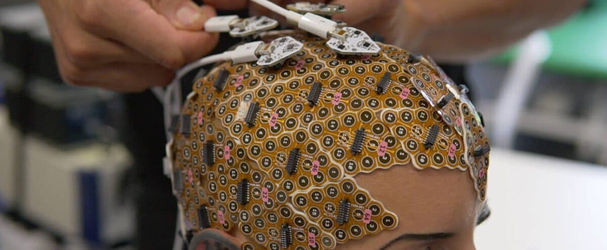
How the Study Was Done
The g.Pangolin 1024-channel EEG system was used to map the central sulcus in five participants through electrical stimulation of the median nerve. An algorithm developed together with Nuri Firat Ince and Dr. Priscella Asman was then applied to locate the central sulcus by detecting the phase reversal of the evoked potentials.
After this, the five participants underwent an MRI scan to verify the position of the central sulcus. The precise agreement between the MRI and EEG data in locating the central sulcus produced truly striking results.
Key Technical Feature
Compared with conventional EEG systems, the g.Pangolin system has far more sensors and therefore a far higher resolution. This makes it possible to obtain much more detailed information about the brain.
Why Does the Central Sulcus Matter So Much?
In neurology, the central sulcus is usually located by placing ECoG grids directly on the brain: the median nerve at the wrist is then stimulated electrically. This produces evoked potentials (EPs) in both the sensory and the motor cortex. A phase reversal appears: an early positive wave over the motor cortex and an early negative wave over the sensory cortex. The central sulcus lies between the ECoG electrode positions that show the negative and the positive responses.
The technique is clinically efficient and can pinpoint this neurosurgical landmark precisely within just a few minutes. But it is an invasive method. This study with g.Pangolin makes it possible to obtain the same results non-invasively.
Visualising Brain Activity with SSEP Classification
Visual representations of brain activity were created using SSEP (somatosensory evoked potential) classification. The results are presented from three different perspectives:
- (a) Ground truth
- Channels in front of the central sulcus are shown in red, those behind it in blue. Grey spheres represent channels with poor signal quality.
- (b) Peak detection
- This map, which shows the intensity of brain activity in colour, highlights the central sulcus much as the reference does.
- (c) Spectral clustering
- A clear separation between the sensory (blue) and motor (red) areas; bad channels are shown in grey.
How Does the g.Pangolin System Work?
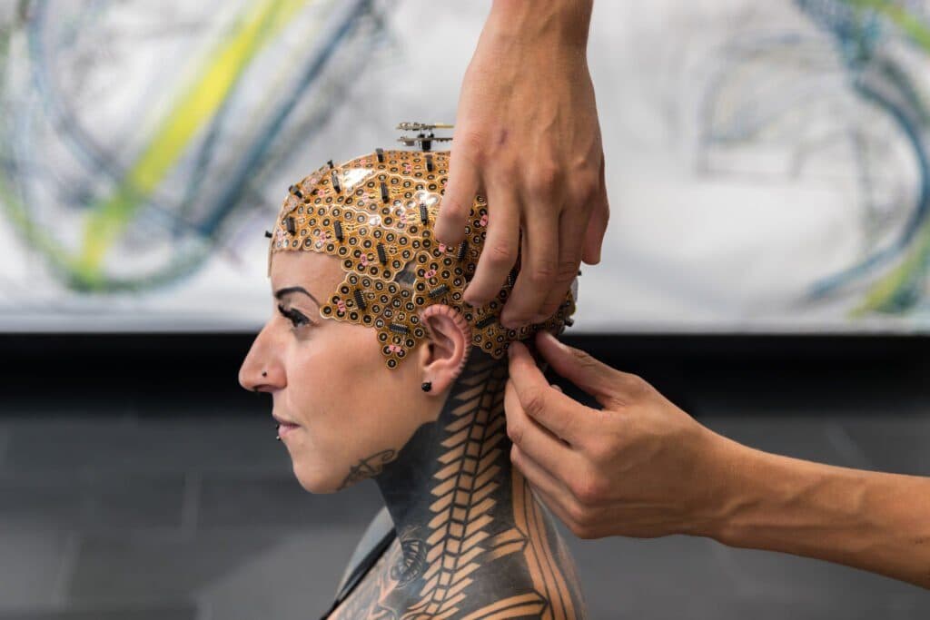
With the g.Pangolin Ultra-High-Density system, participants are prepared by shaving the scalp to make it easier to place the 1024 EEG channels on it. The electrode grids are fixed to the skin with adhesive washers and filled with a special electrode paste to ensure optimal contact and low impedance. A preamplifier is then connected to the electrode grid to record high-quality EEG data.
After electrical stimulation, the evoked potentials are recorded, and an automatic detection algorithm identifies the positive and negative waves, detects the phase reversal and determines the position of the central sulcus.
Technical Set-up
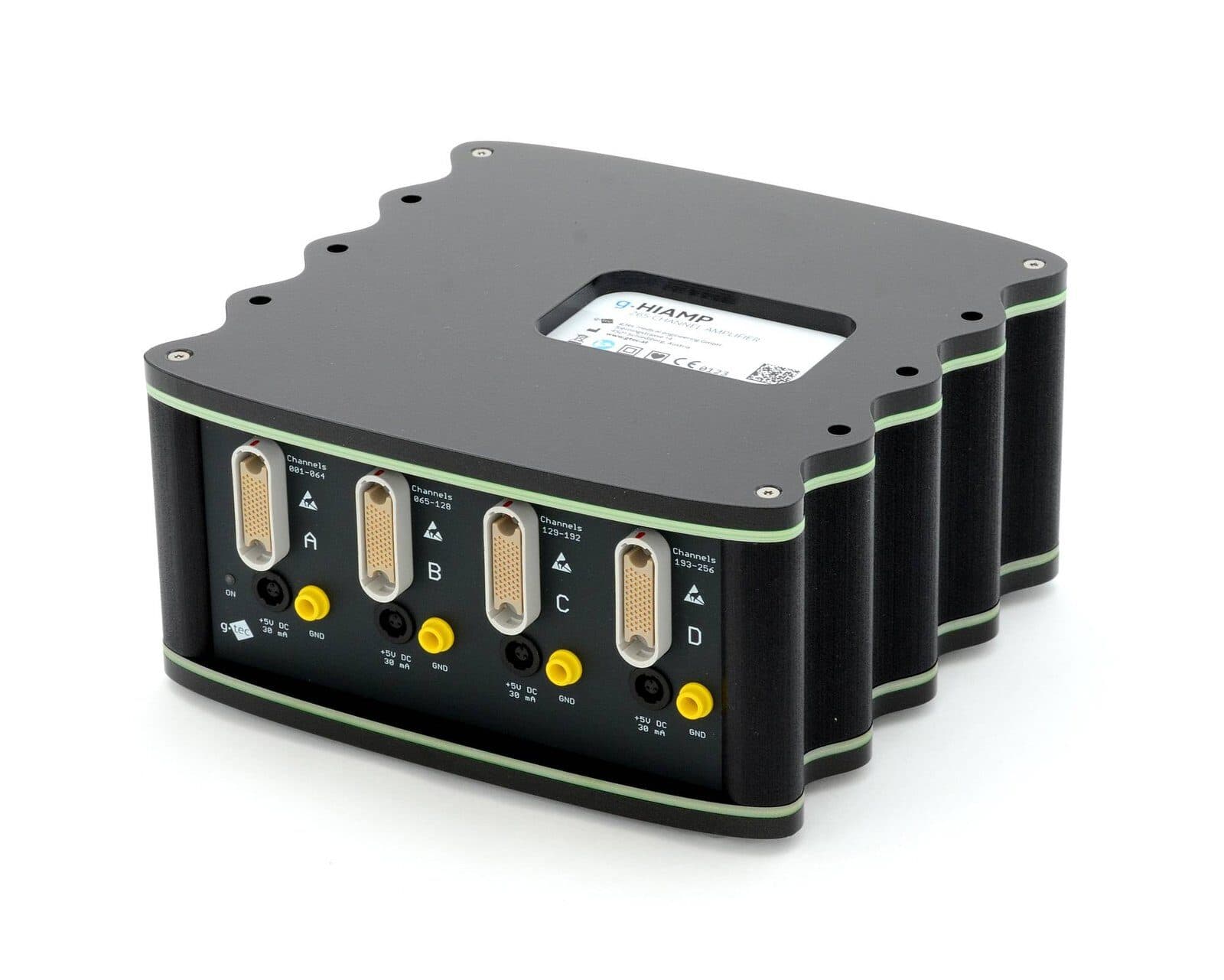
- g.Pangolin electrodes → g.HIamp
- A 256-channel biosignal amplifier with 24-bit resolution. Equipped with four processors, the amplifier has ultra-steep anti-aliasing filtering and high oversampling, and delivers impressive signal-to-noise ratios.
- g.HIsys Professional
- Used for real-time processing, this software is a component of the g.tec Suite 2020 software environment and makes experiments of this kind easier to run.
Results
The study’s findings showed that uHD EEG can locate the central sulcus accurately, with a success rate comparable to that of more invasive methods. This means that we can now understand how different regions of the brain work together without the need for surgery.
Key finding: the precise agreement between the MRI and EEG data in locating the central sulcus is truly striking. This non-invasive approach with g.Pangolin reaches the clinical accuracy obtained with the invasive ECoG method.
Applications
This new approach could be useful in many fields:
- Brain research
- Surgical planning
- Patient monitoring in hospital
- Detecting consciousness
- Controlling a computer by thought (BCI)
- Recovery from injuries
- Improving mental health treatments